She came to me with a report in her hand. The ultrasound had been routine, part of a general health check ordered by her physician. She was 53, two years past her last period, and in good health. And there, at the bottom of the report, was a phrase she had not expected: “simple ovarian cyst, right ovary, 3.2 cm.”
She had not had any pain. No bleeding. Nothing to suggest anything was wrong. But the word “cyst” had sat with her for three days before she made the appointment.
“What does this mean?” she asked. “Should I be worried?”
The short answer is that most postmenopausal ovarian cysts are benign and do not require surgery. The longer answer is that a cyst found in a woman who has gone through menopause is handled differently from one found in a woman who is still menstruating. Not because it is more dangerous, but because the reasons a cyst forms change after menopause, and the evaluation needs to reflect that.
This post explains what a postmenopausal ovarian cyst means, how doctors assess it, and what the different outcomes typically look like.
Why a Postmenopausal Cyst Is Handled Differently
During the reproductive years, the ovaries cycle every month. They grow follicles, release eggs, and form corpus luteum cysts as a normal part of this process. Most cysts in a younger woman are called functional cysts because they are a function of ovulation. They appear and usually disappear within one or two cycles without any treatment.
After menopause, the ovaries stop cycling. There are no follicles being stimulated, no eggs released, no corpus luteum forming. So a cyst found in a postmenopausal ovary is not a functional cyst. It did not form as part of a normal process.
This does not mean it is dangerous. Most postmenopausal cysts turn out to be benign. But because the most common explanation, ovulation, no longer applies, every postmenopausal ovarian cyst is evaluated to understand what it is. The evaluation is a step toward reassurance, not a warning in itself.
The same principle applies to other unexpected gynaecological findings after menopause. If you have had post-menopausal bleeding, or concerns about discharge, the approach is similar: evaluate first, then make decisions based on what is actually found.
Understanding What the Ultrasound Report Is Saying
A pelvic ultrasound (transabdominal or transvaginal) gives the doctor several pieces of information about a cyst. The terminology can look alarming on first reading, but it is mostly descriptive.
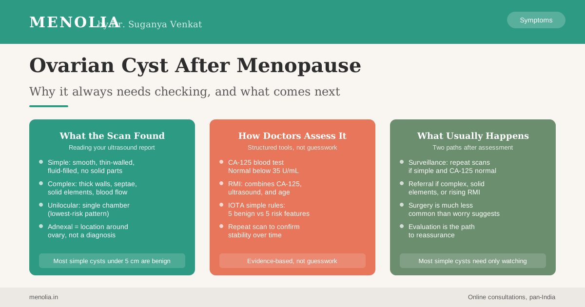
Simple vs complex. A simple cyst appears as a smooth, thin-walled, fluid-filled sac with no internal structures. No solid parts. No septae (internal walls). No detectable blood flow inside. A complex cyst has one or more of these features: thick walls, septae, solid components, calcification, or visible blood flow within the cyst wall or interior. Simple cysts carry a much lower risk than complex ones.
Unilocular vs multilocular. A unilocular cyst is a single chamber. A multilocular cyst has multiple compartments separated by internal walls. Unilocular simple cysts have the lowest risk profile of all postmenopausal cyst types.
Size. Cysts under approximately 5 cm that are simple and unilocular are managed more conservatively than larger ones or those with any complex features.
Adnexal mass or adnexal cyst. “Adnexal” refers to the structures beside and around the uterus, including the ovaries and fallopian tubes. If your report says “adnexal cyst” or “adnexal mass,” that is a location descriptor. It tells you where the finding is, not what it is.
If you are uncertain about the terms on your report, your gynaecologist can walk you through what each finding means for your specific situation.
What CA-125 Shows, and What It Does Not
CA-125 is a protein that can be elevated in certain conditions, including ovarian cancer. A blood test measures its level in circulation. The upper normal limit used in postmenopausal women is generally 35 units per millilitre (U/mL).
It is worth understanding both what CA-125 shows and where its limits are.
CA-125 is not a standalone screening test for ovarian cancer in the general population. Its predictive value in a low-risk, asymptomatic population is limited because the marker can also be elevated in several benign conditions: uterine fibroids, endometriosis, liver disease, inflammatory bowel conditions, and even pelvic infections. A positive result alone does not tell you what is causing the elevation.
Where CA-125 is genuinely useful is as one component of a structured assessment when a cyst has already been found on imaging. Combined with ultrasound features and menopausal status, it contributes to a fuller picture of risk.
If your CA-125 is elevated, it does not mean cancer. It is one piece of information your gynaecologist will use alongside the scan findings to decide the next step.
Practical note for India: CA-125 testing is available at most major pathology networks (Thyrocare, SRL, Dr. Lal PathLabs, Metropolis) in the range of Rs 600 to 1,200 depending on the city and centre. It requires a prescription from your gynaecologist or physician.
How Doctors Assess Risk: RMI and IOTA Simple Rules
Two structured tools are commonly used to bring all the information together.
The Risk of Malignancy Index (RMI) was developed by Jacobs and colleagues (Jacobs I et al., BJOG, 1990; PMID 2223684) and combines three pieces of information: the CA-125 result, the ultrasound score, and menopausal status. A higher RMI suggests higher risk and generally prompts referral to a gynaecological oncologist for further evaluation.
The IOTA Simple Rules (Timmerman D et al., BMJ, 2010; PMID 21156740) use five ultrasound features associated with benign cysts (B-rules) and five features associated with malignant cysts (M-rules) to classify a mass as likely benign, likely malignant, or inconclusive. These rules have been validated across multiple centres and are widely used in specialist settings.
You do not need to apply these tools yourself. Your gynaecologist or the referring specialist will do this. What is useful to understand is that there is a structured, evidence-based process behind the assessment, not a subjective impression.
What Comes Next: Surveillance or Referral
Most postmenopausal cysts, once assessed, follow one of two paths.
Surveillance. For simple, unilocular cysts under approximately 5 cm with a normal CA-125, many gynaecological guidelines support a watching approach: a repeat ultrasound in a few months to confirm the cyst is stable. If it remains unchanged over serial scans, no intervention is usually needed. Regular annual review is part of a sensible post-menopause health plan.
Further assessment or surgical evaluation. Complex cysts, those with solid elements, cysts with an elevated CA-125 or a high RMI score, and cysts that are growing over time are referred to a gynaecologist or gynaecological oncologist for further evaluation. Depending on what is found, this may lead to laparoscopic cyst removal (ovarian cystectomy) or, in some cases, more detailed surgical staging.
The decision about which path is appropriate depends on the specifics of your scan and your blood results. Your gynaecologist will advise you based on your individual findings, not on a generic protocol.
If you have received a scan report showing a cyst and would like to discuss what the findings mean for you, I am available for a video consultation.
Talk to Dr. Suganya on WhatsApp
The Reassuring Reality
It is worth saying clearly: the large majority of postmenopausal ovarian cysts are benign. Studies consistently show that simple unilocular cysts in postmenopausal women, even when they require monitoring, almost always turn out to be cysts with no malignant potential.
Evaluation does not equal danger. The fact that a cyst is being assessed carefully reflects appropriate medical practice, not a high-risk situation. Most of the time, serial ultrasound confirmation of stability is all that is needed. Surgical intervention in postmenopause for a simple cyst is much less common than the worry around these findings might suggest.
Finding a cyst on a scan does not mean something has gone wrong. It means there is a finding that has been noted, as it should be, and that your care team is looking at it systematically.
What to Do Next in India
If a cyst has been found on a pelvic ultrasound, here is what the practical pathway typically looks like.
Your gynaecologist will review the report and discuss whether a CA-125 is needed, whether a repeat scan is appropriate in the short term, or whether a referral to a gynaecological oncologist is warranted. In major Indian cities, gynaecological oncology services are available at Apollo Hospitals, Tata Memorial Centre (Mumbai), Kidwai National Cancer Institute (Bangalore), Rajiv Gandhi Cancer Institute (Delhi), and large government teaching hospitals.
For a simple cyst in someone with no symptoms and a normal CA-125, a watching approach is appropriate and commonly recommended. Your gynaecologist will advise you on the interval for follow-up scans.
If you are unsure about the next steps, it is entirely reasonable to ask your gynaecologist to explain the report in plain language, to clarify what features your cyst has, and to ask what symptoms would prompt you to come back sooner rather than waiting for a scheduled scan.
A cyst found in the course of routine assessment is a different clinical situation from one discovered because of symptoms. Many cysts are found incidentally on scans ordered for entirely different reasons, back pain, urinary symptoms, a routine gynaecological check-up. This is actually a good outcome: identifying something that warrants monitoring, before it causes symptoms.
Frequently Asked Questions
What is a postmenopausal ovarian cyst? A postmenopausal ovarian cyst is a fluid-filled or fluid-and-solid sac found on one or both ovaries in a woman who has completed menopause (12 or more consecutive months without a period). Unlike cysts in the reproductive years, which are usually functional and related to ovulation, postmenopausal cysts form through different mechanisms. Most are benign, but all are evaluated to confirm this.
Does a postmenopausal cyst always need surgery? No. The majority of simple, unilocular cysts under approximately 5 cm with a normal CA-125 are managed with serial ultrasound monitoring rather than surgery. Surgical evaluation is generally reserved for complex cysts, those with solid elements, those with an elevated RMI score, or those that grow on follow-up imaging.
What is CA-125 and what does an elevated result mean? CA-125 is a protein measured in a blood test. It can be elevated in ovarian cancer but also in several benign conditions including fibroids, endometriosis, and liver disease. A result below 35 U/mL is generally considered normal in postmenopausal women, but an elevated result does not mean cancer. It is one piece of information your gynaecologist uses alongside the ultrasound findings to assess the overall picture.
What does “adnexal cyst” mean on an ultrasound report? “Adnexal” refers to the structures around the uterus, including the ovaries and fallopian tubes. “Adnexal cyst” is a location descriptor: it tells you where the cyst is, not what kind it is. Your gynaecologist will review the full report description to determine the nature of the cyst.
How often should I get a follow-up scan? For a simple, low-risk cyst, your gynaecologist will typically advise a repeat ultrasound in three to six months to confirm stability, then annually if it remains unchanged. If the cyst grows or its features change on imaging, the follow-up plan will be adjusted accordingly.
What symptoms should prompt me to contact my doctor sooner? Contact your gynaecologist sooner if you develop pelvic or abdominal pain, a sensation of pressure or heaviness in the lower abdomen, new or unusual bloating, or any urinary changes. These would warrant earlier review rather than waiting for a scheduled scan. Our guide to menopause red flags covers other signs that need prompt attention.
Can ovarian cysts come back after menopause? Yes, a new cyst can form after a previous one has resolved or been removed. This does not signal anything different from a first finding. Each cyst is evaluated on its own features and clinical context. Recurrence is managed the same way: assessment of size, ultrasound character, and CA-125, then a decision about surveillance or further investigation.
A scan finding can feel worrying when you are reading it alone. If you would like to go through your report with me and understand what it means for your health, book a video consultation.
Dr. Suganya Venkat, OB-GYN, Menolia. Online consultations, pan-India, via video call.