Suma had been meaning to get her mammogram for three years. Life kept intervening: her mother’s hip surgery, her daughter’s wedding, her own sense that there was nothing to feel, no lump, nothing to check. At 53, she finally came in for a general review. We organised a mammogram as part of her post-menopausal screening. Her BI-RADS result was 2, which means benign calcifications, a completely normal finding that needs no follow-up beyond routine screening. She felt relieved, of course, but the more lasting feeling was this: she wished she had done it sooner, because the years of postponing had given the fear more room to grow than the mammogram ever warranted.
Most mammogram anxiety comes from the unknown. Women do not know when to start, what the report categories mean, or what happens if something comes back. This post walks through each of those questions, practically and in plain terms.
Why Breast Cancer Screening Matters After 45
Breast cancer is the most common cancer diagnosed in Indian women. According to Global Cancer Statistics 2020 (Sung H, et al., CA Cancer J Clin, 2021, PMID 33538338), it accounts for approximately 26.6 per 100,000 women in India, with incidence rising steadily after 40. Indian women are also often diagnosed at a younger age than their counterparts in Western countries, and are more likely to present with locally advanced disease at first contact.
The reason screening matters is that it detects breast cancer before symptoms appear. A tumour found on a mammogram at 8mm behaves very differently from one that presents as a palpable lump at 3cm. Early detection does not guarantee good outcomes, but it substantially improves the range of treatment options available.
The International Agency for Research on Cancer reviewed the evidence from multiple randomised trials and concluded in their 2015 Handbook on Breast Cancer Screening that invitation to mammography screening reduces breast cancer mortality in women aged 50-69 by approximately 23% among those who attend. The reduction in mortality is the clinical justification for recommending screening at a population level.
When to Start: The Age Question in India
There is no single age threshold that all guidelines agree on, and the Indian picture is complicated by the fact that national guidelines have historically differed from international ones. Here is where the main bodies currently stand.
USPSTF (2024 update): The US Preventive Services Task Force updated its recommendation in April 2024, advising biennial screening mammography for all average-risk women starting at age 40. This was a significant change from the previous recommendation, which had placed the start age at 50. The update was driven by data showing that younger women with breast cancer have better outcomes when the disease is caught early, and that starting at 40 increases the years of potential benefit.
American Cancer Society: Annual mammography from 45 is the ACS recommendation for average-risk women, with the option to start at 40 for those who choose to after discussing benefits and limitations with their doctor. From 55 onwards, women can move to biennial screening if their prior results have been normal.
IARC: The IARC evidence review supports screening in the 50-69 age group for organised population-based programmes. This reflects the evidence base from European randomised trials, where breast cancer age-at-onset patterns differ from the Indian population.
Indian oncology context: Several Indian oncology bodies, including the Indian Cancer Society and the Association of Breast Surgeons of India, have recommended starting screening at 40 for average-risk Indian women, acknowledging the younger age at presentation seen in India. In practice, most major Indian hospitals and oncology centres follow a 40-45 start for average-risk women.
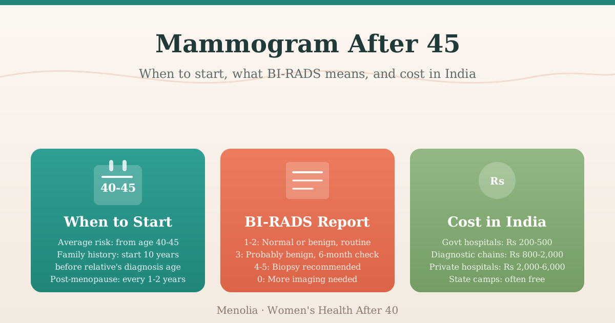
Practical guidance for the post-45 audience: Given all of this, my recommendation is to begin screening by 40-45 if you are at average risk. If you have a first-degree relative (mother, sister, or daughter) who was diagnosed with breast cancer, begin screening 10 years before the age at which your relative was diagnosed, or at 30, whichever is earlier. If you carry a BRCA1 or BRCA2 mutation, or have had prior chest radiation, you are in a high-risk category and will need a different protocol, ideally with guidance from an oncologist.
The post-menopausal years (after 50-52 in most Indian women) are when mammography is most consistently indicated. Oestrogen withdrawal does not directly cause breast cancer, but background risk rises with age, and the post-menopausal years are when the majority of breast cancers are diagnosed in Indian women.
Mammogram or Ultrasound First? Clarifying the Indian Pattern
In India, it is common for doctors and radiologists to order a breast ultrasound before or instead of a mammogram, particularly for women in their 40s. There are reasonable grounds for this, but it is worth understanding the difference between the two tests.
A breast ultrasound is excellent at evaluating a specific area of concern: a lump a doctor has felt, or distinguishing between a solid mass and a fluid-filled cyst. It does not use radiation and it performs better than mammography in women with very dense breast tissue.
A mammogram uses low-dose X-ray to image the entire breast. It is the standard tool for population-level screening because it detects calcifications and architectural changes that are often invisible on ultrasound, and certain patterns of calcification can be an early sign of malignancy.
The two tests are complementary, not interchangeable. For women over 40-45, mammography is the primary screening tool. Ultrasound is used to evaluate something that the mammogram or a clinical examination has flagged. Using ultrasound alone for screening is not the same as getting a mammogram, and the two should not be substituted for each other.
If you have been told to get a breast ultrasound for your annual check, that is reasonable as an additional test in some circumstances, but it does not replace a mammogram after 45.
Reading Your BI-RADS Report
BI-RADS stands for Breast Imaging Reporting and Data System. It is the classification used by radiologists globally to standardise mammography findings. Every mammogram report will include a BI-RADS category. Here is what each one means.
| BI-RADS | Category | What the radiologist found | What happens next |
|---|---|---|---|
| 0 | Incomplete | Additional views or comparison with prior images needed | Return for more imaging |
| 1 | Negative | No abnormality seen | Routine screening on schedule |
| 2 | Benign | Something visible (e.g., calcifications, a cyst) but confirmed as benign | Routine screening on schedule |
| 3 | Probably benign | A finding with less than 2% chance of malignancy | Short-interval follow-up, usually in 6 months |
| 4 | Suspicious | A finding that needs further evaluation; subdivided into 4A (low suspicion), 4B (moderate), 4C (high) | Biopsy recommended |
| 5 | Highly suggestive | Strong imaging features of malignancy | Biopsy recommended |
| 6 | Known malignancy | Already confirmed by prior biopsy | Active treatment underway |
A BI-RADS 1 or 2 is a clean screen. A BI-RADS 3 requires follow-up imaging but is almost always benign on reassessment. It means the radiologist wants to watch that area over the next 6 months, not that they have found cancer. A BI-RADS 4 leads to a biopsy recommendation, which is how a diagnosis is made or definitively ruled out.
If your report says BI-RADS 0, that is not a worrying result. It means the radiologist wants a slightly different view or wants to compare your images with an older study. Call the radiology centre and follow their guidance.
If you have received a mammogram report and are unsure what to make of the BI-RADS result, Dr. Suganya is available for online consultations, pan-India by video call. Message her on WhatsApp at wa.me/919940270499 to book a review.
Dense Breasts: What Your Report Might Say
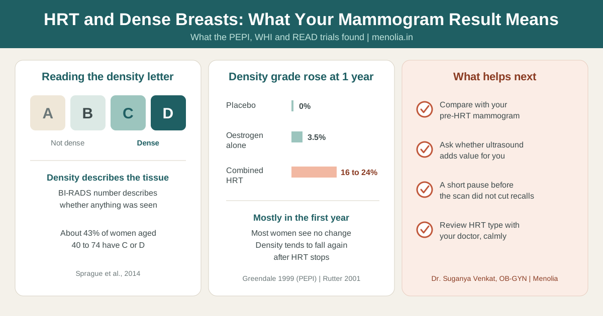
Breast density is assessed on every mammogram and reported in one of four categories: almost entirely fatty (A), scattered areas of fibroglandular density (B), heterogeneously dense (C), or extremely dense (D).
Dense breast tissue appears white on a mammogram, and so does cancer. This means that in women with very dense breasts, a mammogram is less sensitive at detecting small cancers, because a tumour can be obscured against the dense background. Categories C and D are considered dense; A and B are not.
Dense breast tissue is also an independent risk factor for breast cancer, though a modest one.
If your report identifies heterogeneously dense (C) or extremely dense (D) breasts, your doctor may recommend supplemental screening, usually a breast ultrasound alongside the routine mammogram. This is not a reason for alarm; it is an additional layer of safety.
Many perimenopausal women and some post-menopausal women on hormonal therapy have denser breast tissue than their older counterparts. Density typically decreases after menopause as fatty tissue gradually replaces glandular tissue, which is one reason why mammography sensitivity tends to improve in the post-menopausal years.
What a Mammogram Can and Cannot Tell You
A mammogram screens for abnormalities that warrant further evaluation. It does not diagnose cancer by itself. A BI-RADS 4 or 5 result requires a biopsy for confirmation; the mammogram alone is not sufficient for a cancer diagnosis.
What mammography detects well: microcalcifications (tiny calcium deposits that can be an early sign of certain cancers, including ductal carcinoma in situ), masses or architectural distortions, and asymmetries between the two breasts.
What it detects less reliably: cancers in very dense breast tissue, and some forms of lobular carcinoma in early stages.
The goal of screening mammography is to find changes at a stage when treatment is less extensive and outcomes are better. It will not find every cancer, and it does produce a proportion of false-positive results. Most BI-RADS 3 and 4A findings turn out to be benign on follow-up. A recall for additional imaging is far more likely to end with reassurance than with a diagnosis.
Cost of a Mammogram in India
Costs vary significantly depending on the setting. The figures below are approximate and verified as of mid-2026.
| Setting | Approximate cost |
|---|---|
| Government hospitals (district, medical college) | Rs 200 to 500 (subsidised; availability varies by state) |
| CGHS-empanelled diagnostic labs | Rs 400 to 800 (for central government employees and their families) |
| Diagnostic chains (SRL, Thyrocare, Neuberg Diagnostics) | Rs 800 to 2,000 |
| Mid-range private hospitals | Rs 2,000 to 4,000 |
| Tertiary private hospitals (Apollo, Fortis, Manipal) | Rs 3,500 to 6,000 |
| State cancer screening camps | Often free on National Cancer Awareness Day and during state scheme camps |
Digital mammography, which produces higher-resolution images and allows radiologist-to-radiologist sharing, is now available at most chain diagnostics and major private hospitals across India. If you have had a prior mammogram elsewhere, bring those images or request the old films, because comparison with previous studies is an important part of interpreting the new report.
Tamil Nadu’s Innuyir Kappom Thittam (Chief Minister’s Comprehensive Health Insurance Scheme) and similar state-level cancer screening initiatives include breast screening in their programme components. Ask at your nearest government hospital or PHC about camp schedules.
Self-Examination: Useful, but Not a Replacement
Monthly breast self-examination has been taught for decades, and it remains useful for one specific purpose: helping you learn what your breasts normally feel like, so that you notice when something changes. A Cochrane systematic review by Kosters and Gotzsche (2003, CD003373, PMID 12804462) examined two large randomised trials and found that breast self-examination did not reduce breast cancer mortality. Women who practised it had more benign biopsies but no better survival outcomes than those who did not.
A lump or change you find while examining yourself is a reason to see a doctor promptly. Prompt reporting matters, and formal imaging should still follow.
Symptoms that warrant prompt review, regardless of your next scheduled mammogram:
- A new lump or thickening in the breast or armpit
- Skin changes on the breast: redness, dimpling, puckering, or a peau d’orange (orange-peel) texture
- Nipple changes: inversion that is new, scaling, or discharge, particularly blood-stained discharge
- A change in breast size or shape that appears without explanation
- Persistent, localised breast pain in one spot (though most breast pain in perimenopause is hormonal and not associated with cancer)
For more on what hormonal breast tenderness in perimenopause looks like and how it differs from pain that needs evaluation, read our post on menopause and breast tenderness.
Putting Together Your Screening Plan After 45
If you are between 40 and 45 and have not had a mammogram: this is a reasonable time to start, particularly if you have any family history or personal risk factors. Ask your gynaecologist at your next consultation.
If you are between 45 and 55: this is the age range where screening is most consistently recommended across all major guidelines. There is no clinical reason to delay.
If you are past 55 and have not had a mammogram in two or more years: organise one. Post-menopausal screening continues to be valuable, particularly because oestrogen is no longer masking early symptoms in the way it sometimes does in the reproductive years.
If you are on HRT: this does not change the mammogram recommendation. HRT does not disqualify you from screening, and it does not change the frequency. The relationship between HRT and breast cancer risk is a separate, nuanced discussion. For a detailed look at how HRT decisions are made, read our post on HRT in India: what an OB-GYN actually recommends.
Mammography is one part of post-menopausal screening. For the full picture, including bone density, cholesterol, blood sugar, and thyroid, read our post-menopause health checks guide. If you are also thinking about colorectal cancer screening, which is now recommended from 45, our colorectal cancer screening guide covers the FIT test versus colonoscopy decision in detail. For bone density, see our DEXA scan and bone density test guide.
The three tests together, a mammogram, a DEXA scan, and a colonoscopy or FIT test, form the core of preventive screening in the post-menopausal years. They are regularly delayed because they feel elective in the absence of symptoms. They are not elective. They are how problems are found before they become crises.
Frequently Asked Questions About Mammograms After 45
At what age should Indian women get their first mammogram?
Most Indian oncology bodies recommend starting at 40-45 for average-risk women. The 2024 USPSTF update recommends 40 for all average-risk women. If a first-degree relative was diagnosed with breast cancer, start 10 years before her diagnosis age, or at 30, whichever comes first. There is no single national Indian guideline with one fixed age, but 40-45 is the practical consensus for average-risk women.
How often should I get a mammogram?
Once a year is the most widely recommended frequency for women aged 40-54 at average risk. From 55 onwards, every two years is considered acceptable if prior screens have been normal. Women with dense breasts, a personal history of breast abnormalities, or a family history may be advised to continue annual screening regardless of age. Discuss frequency with your gynaecologist based on your specific situation.
My report says BI-RADS 3. Should I be worried?
BI-RADS 3 means the finding is probably benign, with less than 2% likelihood of being malignant. It does not mean cancer has been found or is suspected. It means the radiologist wants to recheck that area in 6 months to confirm the finding is stable. At 6-month follow-up, the large majority of BI-RADS 3 findings are reclassified as BI-RADS 2. A BI-RADS 3 recall is a precaution, not a diagnosis.
My report says I have dense breasts. What should I do?
Dense breast tissue makes mammography less sensitive because both dense tissue and early cancers appear white on the X-ray. If your report identifies heterogeneously dense (C) or extremely dense (D) breasts, your doctor may recommend a breast ultrasound alongside your routine mammogram. Dense breasts are a normal finding, not a disease. They are more common in younger women and in those on hormonal therapy. Density often decreases after menopause as glandular tissue is gradually replaced by fatty tissue.
I have no family history of breast cancer. Do I still need a mammogram?
Yes. Approximately 70-80% of breast cancers occur in women with no family history of the condition. Family history raises your risk, but its absence does not mean you are protected. Mammography is a population-level screening tool, recommended for all women over 40-45 regardless of family history.
What is the difference between a breast ultrasound and a mammogram?
A breast ultrasound uses sound waves and is excellent for evaluating a specific area of concern, distinguishing a solid mass from a cyst, or assessing women with very dense breast tissue. A mammogram uses low-dose X-ray to image the entire breast and is the standard tool for population-level screening. Ultrasound does not detect microcalcifications, which are among the most important early mammographic findings. After 45, a mammogram is the primary screening test. Ultrasound may complement it in certain situations, but it does not replace it.
My mammogram showed calcifications. Is that something to worry about?
Calcifications are calcium deposits in breast tissue. They are extremely common and are almost always benign. Large, coarse macro-calcifications are virtually always a BI-RADS 2 finding. Tiny microcalcifications, particularly in certain cluster patterns or linear arrangements, are the type that radiologists examine carefully, because specific patterns can be an early sign of ductal carcinoma in situ. If your radiologist assigns BI-RADS 3 or 4, they want follow-up imaging or a biopsy to characterise those calcifications more precisely. Most such findings turn out to be benign on further evaluation.
The appointment takes approximately 20-30 minutes. The report is usually available within 24-48 hours. Most women walk away with a BI-RADS 1 or 2 and nothing to follow up.
If you have never had a mammogram, or your last was more than two years ago, this is the prompt to book it. If you would like to discuss your personal screening timeline, understand a report you have received, or build a broader post-menopausal health plan, Dr. Suganya is available for an online consultation by video call, pan-India. Message her on WhatsApp at wa.me/919940270499 to book your consultation.